Expression of molecular markers in mouse and human stellate ganglia
We used immunohistochemistry to visualize the expression of molecular markers in stellate ganglia from mice and human. Data consist of images (CZI format) of SG cryosections stained with antibodies combinations, acquired with a Zeiss 710 microscope.
Dataset Overview
Study Purpose: Sympathetic regulation of peripheral organ function is achieved through the coordinated actions of secretomotor, vasomotor and erector sympathetic neurons, in a manner that is highly conserved across species. Functionally distinct classes of sympathetic neurons have been defined by their firing properties and the expression of different combinations of neurotransmitters and neuropeptides. The identification of unique molecular markers for these populations would open the door to the application of genetic tools to parse the actions of secretomotor, vasomotor and erector sympathetic neurons. We identified unique molecular markers that can distinguish between subpopulations of stellate ganglion (SG) neurons in the mouse. Here we investigated whether the expression patterns of these markers are conserved in humans by performing immunohistochemistry (IHC) on SG cryosections.
Data Collection: Data consists of 5 tilescan images (CZI format) of human and mouse SG using a Zeiss LSM 710 inverted confocal microscope.
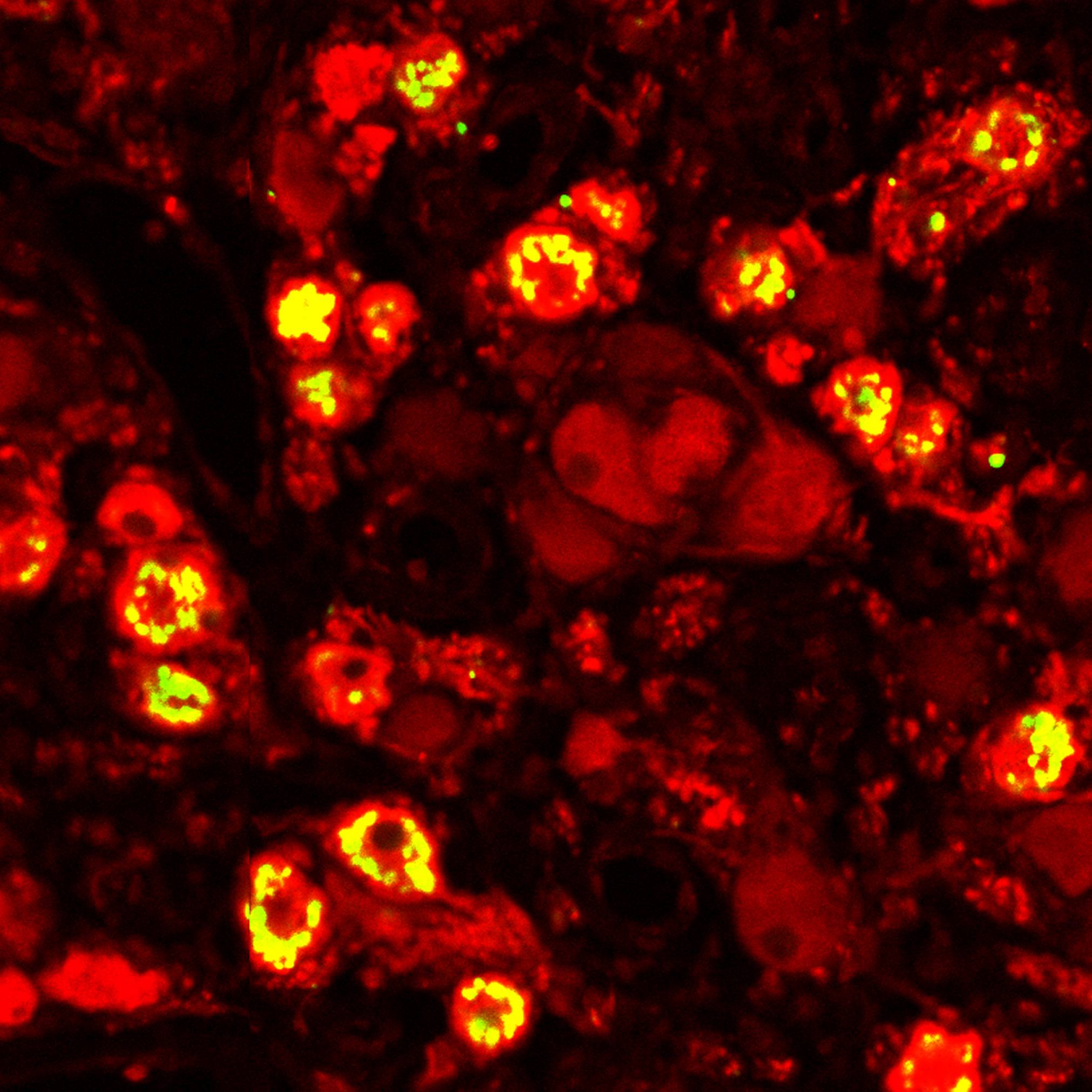
Primary Conclusion: Most commercial antibodies tested did not provide reliable results. We were able to clearly visualize neuropeptide Y (NPY) expression in a subset of tyrosine hydroxylase-expressing (TH+) neurons, while vasoactive intestinal peptide (VIP) expression was complementary to TH.
Curator's Notes
Experimental Design: Both mouse and human stellate ganglia (SG) were fixed in 4% PFA in 0.1M PB overnight at 4°C, washed with Phosphate Buffer Saline (PBS) 4°C for at least 1 h. Ganglia were encased in Optimal Cutting Temperature compound (Tissue-Tek), cut into 10 μm cryo-sections on slides, and stored at -80°C. Tissue sections were subjected to the standard staining procedure with antibody combinations. Expression patterns were visualized by confocal microscopy.
Completeness: This dataset is a part of a larger study, "Expression of molecular markers for SG neuronal subpopulations in 3 sympathetic ganglia."
Subjects & Samples: One adult male mouse (RRID:IMSR_JAX:000664) and one human subject were used in this study. The human SG was kindly provided by Dr. Olujimi Ajijola at UCLA, it was dissected from a 44-year-old female donor.
Primary vs derivative data: Primary data is organized by the subject ID folders and sample ID subfolder. Each sample subfolder contains confocal images (CZI format). Image data in the derivative folder (JPEG2000 and OME-TIFF) was derived from primary images (.CZI).
Files
1 - 0 of 0 files
About this dataset
Publishing history
Cite this dataset
Tags
References
Is Supplemented by
Neri, D. (2022). Expression of molecular markers in mouse and human stellate ganglia v1. https://doi.org/10.17504/protocols.io.rm7vzyko5lx1/v1

Copyright © 2026 University of Pennsylvania. All rights reserved.