Assessment of gastric emptying and motility with magnetic resonance imaging (MRI) under gastric electrical stimulation (GES) in rats
This study aims to evaluate the effects of gastric electrical stimulation (GES) on gastric emptying and motility in rats.
Dataset Overview
Study Purpose: This study aims to evaluate the effects of gastric electrical stimulation (GES) on gastric emptying and motility in rats.
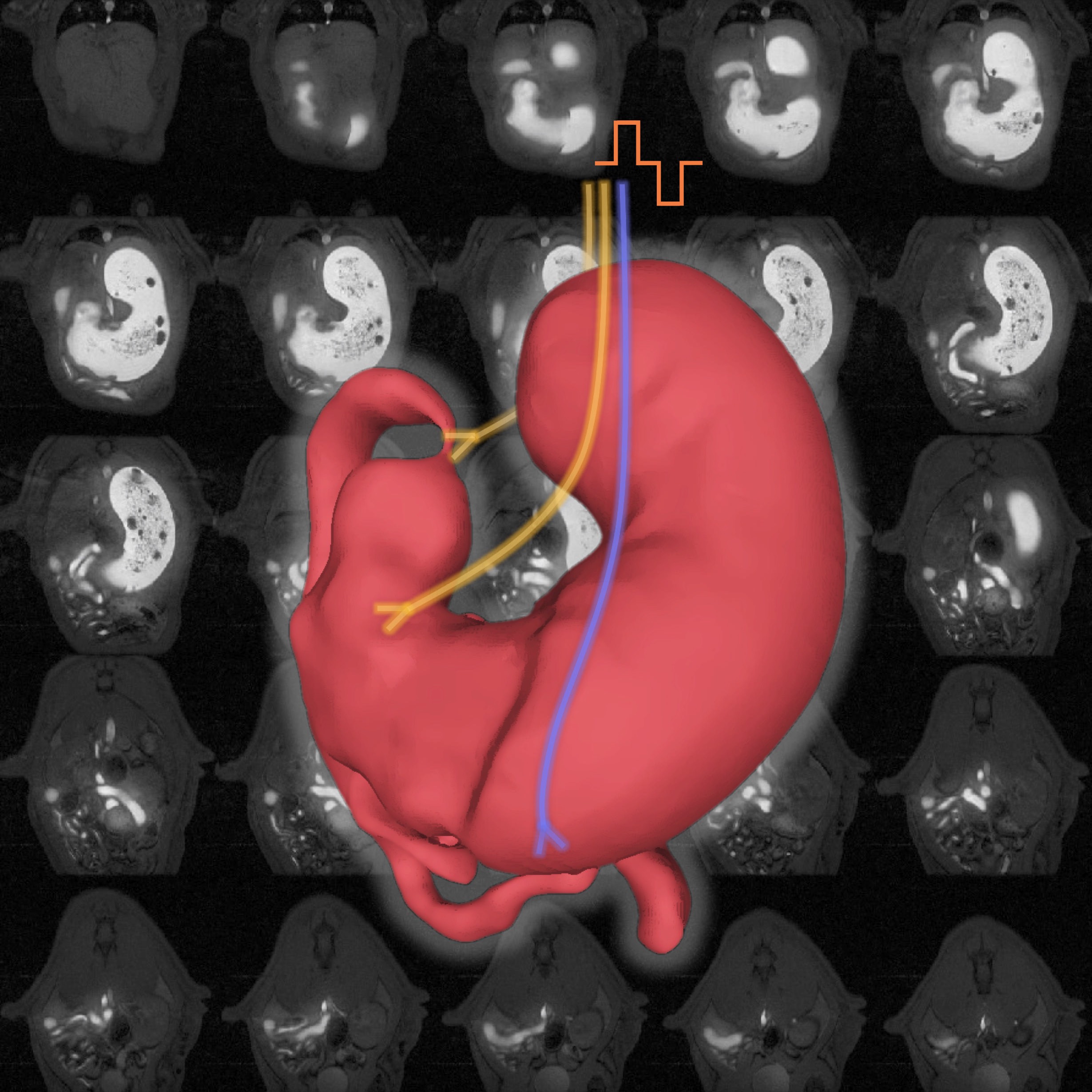
Data Collection: Briefly, a gadolinium‐based contrast agent was mixed with the animal’s meal in order for chyme to appear “bright” in MRI scans, thereby delineating the gastric and intestinal volume. A multi‐slice MRI sequence was used to scan gastrointestinal (GI) functions and physiology with high spatial and temporal resolution.
Primary Conclusion: none stated
Curator's Notes
Experimental Design: Four experiments were performed in this study. In the first study, researchers tested the brain response to low dose gastric electrical stimulation (GES; current = 0.6 mA, pulse width = 0.2 ms, frequency = 5Hz) which followed a 20s-ON-40s-OFF stimulation pattern. In the second study, researchers switched to high dose GES (current = 6 mA, pulse width = 0.3 ms, frequency = 20 Hz) which followed a 2s-ON-3s-OFF stimulation pattern. In the third and fourth study, they performed bilateral cervical vagotomy for each animal and repeated the simulation described in the first and second study, respectively. This dataset reports on animals that were divided into three groups: sham control group including the rats that only received sham surgery (n = 5) and two GES groups including the rats that received both surgery and vagus nerve stimulation (VNS). GES1 stimulated by 0.3mA/0.2ms/10Hz (n=2) and GES2 stimulated by 0.6mA/0.2ms/5Hz (n=4).
Paradigm control / sham surgery
- GES (low); current = 0.6 mA, pulse width = 0.2 ms, frequency = 5Hz
- GES (high); current = 6 mA, pulse width = 0.3 ms, frequency = 20 Hz
surgery + vagus nerve stimulation
- GES1 (v.low); current = 0.3mA, pulse width = 0.2ms, frequency = 10Hz (n=2)
- GES2 (low); current = 0.6mA, pulse width = 0.2 ms, frequency = 5Hz (n=4)
Completeness: This is a part of a larger study: Effects of Vagus Nerve Stimulation/Gastric Electrical Stimulation on Gastric Emptying and Motility Assessed with Magnetic Resonance Imaging
Subjects & Samples: This study used adult Sprague-Dawley male rats (n=11) between 9-11 weeks old.
Primary vs derivative data: Primary data is organized by subject name. Each folder contains data on 3D + time volumetric MRI data along with acquisition time of each 3D volume relative to the first volume (minute), and Nth 3D + time motility MRI data interleaved with volumetric MRI data along with acquisition time of each 3D volume (seconds). Derivative data is also organized by subject name. Each subject folder contains segmented 3D + time volumetric MRI data, and segmentation of stomach, intestine, stomach compartments, antrum, and antral motility map for each experimental subject.
Files
1 - 0 of 0 files
About this dataset
Publishing history
Cite this dataset
Tags
References
Is Supplemented by
Lu, K.-H., Liu, Z., & Cao, J. (2019). Effects of Vagus Nerve Stimulation/Gastric Electrical Stimulation on Gastric Emptying and Motility Assessed with Magnetic Resonance Imaging v1. https://doi.org/10.17504/protocols.io.bawfifbn

Copyright © 2026 University of Pennsylvania. All rights reserved.