Calcium imaging and motility tracking of distinct myenteric neuronal subsets in mice
Calcium imaging videos of mouse enteric neurons
Dataset Overview
Study Purpose: Collaboration between Duke University and the University of Nevada, Reno for computational analysis of myenteric neuronal calcium imaging.
Data Collection: Imaging and stimulating enteric neurons in the murine large intestine.
Primary Conclusion: None stated.
Curator's Notes
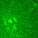
Experimental Design: Adult mice of each sex were killed by isoflurane inhalation, and cervical dislocation, in accordance with the requirements of the Animal Ethics Committee at the University of Nevada, Reno. A ventral midline incision was made, and the entire colon was removed and placed in a Sylgard-lined dish that was perfused with Krebs-Ringer buffer solution. The region of interest was carefully pinned mucosal side up and a camel-hair paintbrush was used for mucosal stimulation, where each stimulation consisted of a sequence of five brush strokes in approximately 5 seconds. Electronic field stimulation was applied at 20 hertz for 10 seconds; 6 volts .01 millisecond pulse duration. Functional imaging was performed on a Nikon Eclipse FN1 upright fluorescence microscope using Nikon Plan Fluor 20x lens. Image sequences were captured using a Photometrics Prime 95B sCMOS camera and captured on a Windows-based PC using Nikon NIS Elements 4.1. Image sequences were recorded at 25 frames per second.
Completeness: This dataset is complete.
Subjects & Samples: Male (n=7) and female (n=8) adult transgenic mice were used in this study.
Primary vs derivative data: The primary data is organized in folders by subject ID. Each sample subfolder contains .tif microscope imaging files of calcium transient and movement responses of cholinergic and nitrergic neurons in the middle colon of male and female mice to oral or anal mucosal or electrical field stimulation. Each imaging file is accompanied by a set of supportive files where ROI is saved as text to make the trace as a xy coordinate file (.xlsx). There is no derivative data folder.
Files
1 - 0 of 0 files
About this dataset
Publishing history
Cite this dataset
Tags
References
Is Supplemented by
Heredia, D., Gould, T., & K Smith, T. (2019). Imaging and stimulating enteric neurons in the murine large intestine v1. https://doi.org/10.17504/protocols.io.82fhybn

Copyright © 2026 University of Pennsylvania. All rights reserved.