Evaluating spheres of influence for efferent neural control of the heart
Evaluating spheres of influence for efferent neural control of the heart In a minipig model, sequential knockdown of cardiac ganglionated plexi (GPs) was performed to cause loss of function.
Dataset Overview
Study Purpose: To evaluate spheres of influence for efferent neural control of the heart.
Data Collection: This dataset includes cardiac electrophysiologic and hemodynamic data with autonomic stimulation before and after two sets of sequential ablations of GPs were performed.
Primary Conclusion: None stated.
Curator's Notes
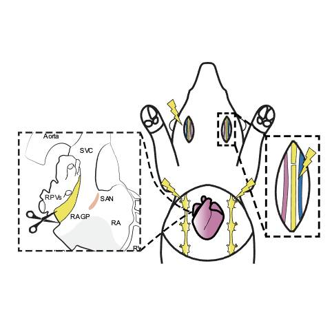
Experimental Design: In a Yucatan minipig model, sequential knockdown of cardiac ganglionated plexi (GPs) was performed to cause loss of function. Disruption of specific GPs of the intrinsic cardiac nervous system (ICN) was performed using an ultrasonic aspirator to define specific areas of influence with respect to neural control of cardiac mechanical and electrical function. Electrical autonomic (parasympathetic and sympathetic) stimulation were the primary methods of assessment for areas influenced by specific ICN ganglia as defined by regional loss of synchrony (electrical) following each successive GP ablation. GPs targeted include the right atrial GP (RAGP), posterior atrial GP (PAGP), inferior vena cava-inferior atrial GP (IVC-IAGP), left atrial GP (LAGP) and ventral intraventricular GP (VIVGP). Cardiac electrical activity was measured using a 56-electrode sock overlying the heart, and a pressure transducer catheter in the heart measured left ventricular pressures.
Completeness: This dataset is a part of a larger study: "Innervation and neuronal control of the mammalian sinoatrial node."
Subjects & Samples: Male (n=7) and female (n=2) Yucatan minipigs (RRID:NSRRC_0012) 9-11 months old were used in this study.
Primary vs derivative data: Primary data is organized in folders by the subject ID and the type of performance. Each performance subfolder contains hemodynamics or cardiac electrophysiology data collected in Spike2 (.txt files) and using the GE CardioLab system (.txt files). There is no derivative data folder.
Files
1 - 0 of 0 files
About this dataset
Publishing history
Cite this dataset
Tags
References
Described by
Hanna, P., Dacey, M. J., Brennan, J., Moss, A., Robbins, S., Achanta, S., Biscola, N. P., Swid, M. A., Rajendran, P. S., Mori, S., Hadaya, J. E., Smith, E. H., Peirce, S. G., Chen, J., Havton, L. A., Cheng, Z. (Jack), Vadigepalli, R., Schwaber, J., Lux, R. L., … Shivkumar, K. (2021). Innervation and Neuronal Control of the Mammalian Sinoatrial Node a Comprehensive Atlas. Circulation Research, 128(9), 1279–1296. https://doi.org/10.1161/circresaha.120.318458
Is Supplemented by
Hanna, P., Ardell, J., & Shivkumar, K. (2021). Evaluating intrinsic cardiac neural control of cardiac function using sequential ganglionated plexus ablations v1. https://doi.org/10.17504/protocols.io.bvpbn5in

Copyright © 2026 University of Pennsylvania. All rights reserved.