Imaging colon and bladder sensory convergence in CLARITY cleared mouse spinal cord
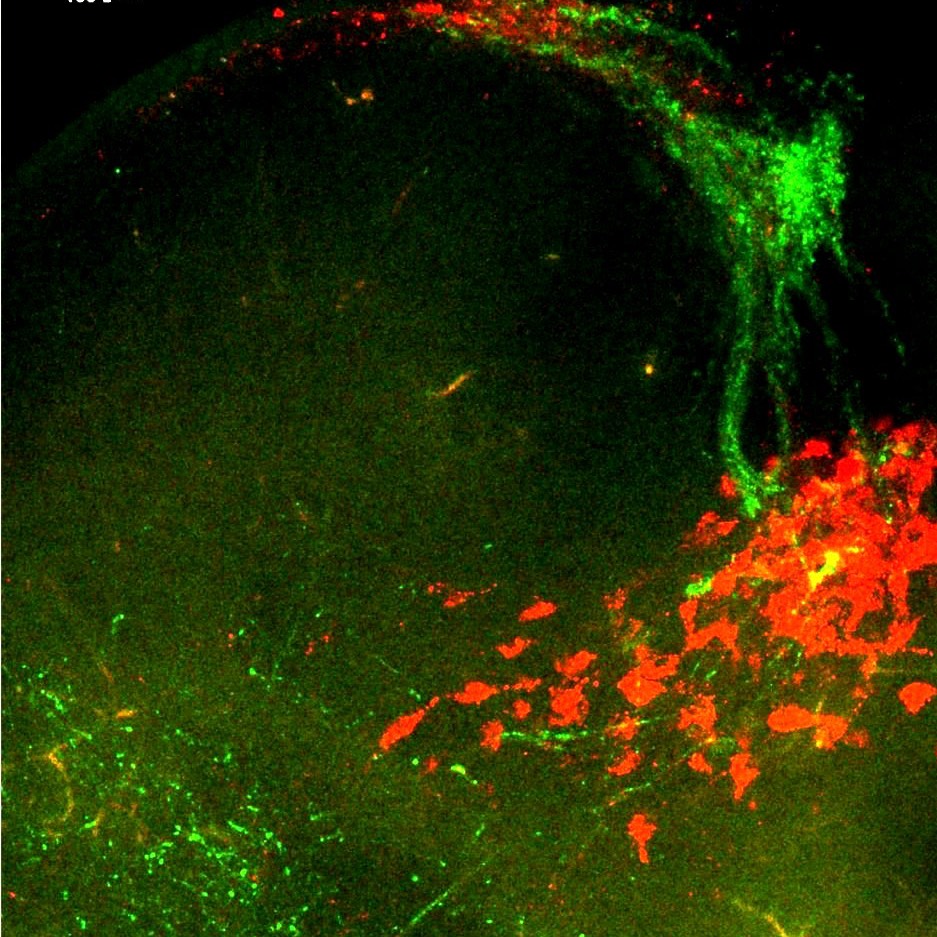
Colon and bladder innervating sensory nerves were identified by retrograde tracing and imaged in the whole CLARITY cleared spinal cord to identify the distribution of colon/bladder dichotomizing input.
Dataset Overview
Study Purpose: Identify the distribution of colon/bladder sensory nerve input in the mouse spinal cord.
Data Collection: Confocal laser scanning microscope (Leica TCS SP8X) was used to optically section cleared lumbosacral spinal cord. Confocal images (1024 × 1024 pixels) were obtained with PL APO CS2 air 10X. Sequential scanning (5 line average) was performed with the following settings using a tunable white light laser and photomultiplier detectors: 495 nm excitation and 503 / 538 nm-emission detection for AF488 and 561 nm-excitation and 570-625 nm-emission detection for AF594. Spinal cords with DRG attached were optically sectioned (5µm thick), and z-projected images were reconstructed for each sample (230-390µm).
Primary Conclusion: The ability to image the intact spinal cord enabled visualization of the spinal regions in which sensory nerves from the colon and bladder project in relation to their sensory neurons located in the DRG. Dense colon and bladder sensory afferent input is localized within discrete regions of the L5-S1 levels of the spinal cord. In the thoracic spinal levels input from colon and bladder, sensory afferents is less abundant. In both spinal levels, colon and bladder dichotomizing sensory afferent input is rare however colon and bladder sensory input into the spinal cord is highlighly convergent in discrete regions.
Curator's Notes
Experimental Design: Mice underwent retrograde tracing using cholera toxin subunit b (ctb) from the colon (ctb-af594) and bladder (ctb-af488) wall. Seven days later, mice underwent transcardial perfuse fixation, and segments of the spinal cord corresponding to thoracolumbar (T9-L2) and lumbosacral (L3-S4) spinal levels were removed with DRG attached and underwent clarity clearing. ctb labelled fibres in the spinal cord and neurons in the DRG were visualized using confocal microscopy, and the distribution of colon and bladder sensory input was qualitatively assessed. n=4 (1m:3f)
Completeness: This dataset is part of a larger study called "Colon/Blader sensory neurons ganglia in the mouse". Related datasets are available.
Subjects & Samples: Male (n=1) and female (n=3) adult mice (RRID:IMSR_JAX:000664) were used in this study.
Primary vs derivative data: Primary data folder contains lumbosacral and thoracolumbar spinal cord Z-stack confocal images as microscopy LEICA files (.lif). The images are organized in folders by the subject name and sample name, respectively. Image data (JPEG2000 and OME-TIFF) was derived from primary images (.lif). Primary images were converted with 40:1 compression to JPEG2000 (.jpx) by MBF Bioscience for web streaming and visualization on the SPARC Data Portal. Primary images were also converted with lossless compression to OME-TIFF (.tif) by MBF Bioscience.
Files
1 - 0 of 0 files
About this dataset
Publishing history
Cite this dataset
Tags
References
Is Supplemented by
Harrington, A. (2022). Mouse model of post-colitis (DNBS) chronic visceral hypersensitivity. v1. https://doi.org/10.17504/protocols.io.14egn7mpqv5d/v1
Harrington, A. (2022). Cholera Toxin Subunit B (CTB) Retrograde tracing from the mouse colon and bladder wall. v1. https://doi.org/10.17504/protocols.io.x54v9y391g3e/v1

Copyright © 2026 University of Pennsylvania. All rights reserved.