Temporal dispersion in porcine subdiaphragmatic nerves ex vivo
This dataset includes ex vivo experimental data on porcine subdiaphragmatic nerves. The data was used for evaluation of the approach for overcoming temporal dispersion in unmyelinated neves designed in the previously performed modelling study.
Dataset Overview
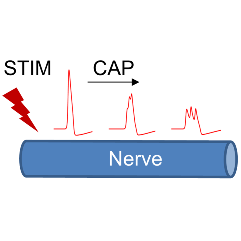
Study Purpose: Fast neural Electrical Impedance Tomography (FnEIT) is an imaging technique that has been successful in visualizing electrically evoked activity of myelinated fibers in peripheral nerves by measurement of the impedance changes (dZ) accompanying excitation. However, imaging of unmyelinated fibers is challenging due to temporal dispersion (TP), which occurs due to variability in conduction velocities of the fibers and leads to a decrease of the signal below the noise with distance from the stimulus. To overcome TP and allow EIT imaging in unmyelinated nerves, a new experimental and signal processing paradigm is required allowing dZ measurement further from the site of stimulation than compound neural activity is visible. The development of such a paradigm was the main objective of this study.
Data Collection: We used an ex vivo preparation of porcine subdiaphragmatic nerves with four multielectrode cuffs. Each studied nerve was subjected to 10 Hz trains of stimuli lasting 0.6 s (with 5 s between trains) applied to a pair of electrodes on the first cuff, and AC current was applied at a range of frequencies through a pair of electrodes on the third or fourth cuff placed approximately 15 and 20 cm from the first one. Voltages were recorded on all electrodes with respect to the last electrode on the last cuff. Each recording lasted 30 minutes.
Primary Conclusion: n = 18 out of 28 recordings had a satisfactory level of noise smaller than 4 μV RMS before averaging, in agreement with the model described in the paper accompanying this dataset. The SNR at 15 cm after 30 minutes of averaging was 1.8 ± 0.7, decreasing to 1.7 ± 0.6 at 20 cm, which is in agreement with the predictions of the developed model. The mean absolute value of the determined dZ across all nerves was found to be significantly larger than the mean at every other point in the recording (p < 0.01, n = 18).
Curator's Notes
Experimental Design: All experimental procedures complied with regulations in the UK Animal (Scientific Procedures) Act, 1986, and were reviewed and approved by the Animal Welfare and Ethical Review Board. For experimental evaluation of the developed method, an ex-vivo setup with the pig's subdiaphragmatic nerves (SN) was used. A detailed description of the experimental setup and the results are provided in the paper accompanying this dataset (https://doi.org/10.1088/1741-2552/ac669a). Porcine SNs were held in an organ bath perfusion chamber filled with continuously oxygenated saline solution. Three silicone rubber cuffs each having six radially arranged electrodes made from stainless steel and coated with PEDOT:pTS were placed around the nerve 3, 15 & 20 cm from the same cuff used for electrical stimulation (fstim= 2 Hz, Istim = 20-40 mA, PW = 50 μs). Impedance changes were measured by sequential application of the sinusoidal current through two last electrodes on each cuff, and the voltage was recorded on the remaining electrodes on the same cuff in respect to the last electrode on the last cuff. Then, dZ was obtained by demodulation of the recorded voltage using the absolute of the Hilbert transform. The applied sinusoidal current parameters were: fAC= 1–6 kHz, IAC= 200-300 μA. The optimal stimulation paradigm for recording dispersed dZ determined with the model was applied to n = 28 nerves to sequentially record dZ using cuffs 3 and 4 at 15 and 20 cm from the onset, where caps were dispersed and not measurable. Signal to noise ratios, SNRs, at these distances were obtained; implications for imaging unmyelinated nerves with FnEIT were investigated. Statistical significance of the recorded dZ was verified using a two-sample t-test algorithm by comparison of the measurement amplitude straight after the stimulation artefacts (600-1000 ms) with the dZ at all other points following this period (1000-3000 ms). The dataset contains N=18 samples in each experimental group (cuff 3 and cuff 4, located at 15 and 20 cm from the stimulating cuff, respectively).
Completeness: This dataset is complete.
Subjects & Samples: Adult female (n-18) pigs, 60-70 kg, were used in this study.
Primary vs derivative data: The data for each sample includes three files: 1) '.eeg'- includes the eeg-type data; 2) '.vhdr'- includes header information for each file; 3) '.vmrk'- includes marker information such as stimulation markers. There is no derivative data folder.
Code Availability: Code for data processing is located in the 'Code' folder. It includes two files:
'post_processing_trains_clean.m' Use this Matlab code for processing the raw data. The code does the following:
i. cutting the signal according to the locations of the triggers, ii. band-pass filtering of the signal around the central frequency with the bandwidth of 100 Hz, iii. demodulation of the signal using the modulus of Hilbert transform to extract impedance change.
'plot_traindZ_clean.m'. This file is used for plotting CAPs and dZs of the processed data. The code does the following:
i. High-pass filtering to reduce low-frequency noise, ii. Subsequent low-pass filtering to reduce the effect of stimulation artefacts and high-frequency noise.
Files
1 - 0 of 0 files
About this dataset
Publishing history
Cite this dataset
Tags
References
Described by
Tarotin, I., Mastitskaya, S., Ravagli, E., Perkins, J. D., Holder, D., & Aristovich, K. (2022). Overcoming temporal dispersion for measurement of activity-related impedance changes in unmyelinated nerves. Journal of Neural Engineering, 19(2), 26054. https://doi.org/10.1088/1741-2552/ac669a
Is Supplemented by
Tarotin, I., Mastitskaya, S., Ravagli, E., D Perkins, J., S Holder, D., & Aristovich, K. (2022). Measurement of activity-related impedance changes in porcine subdiaphragmatic nerve v2. https://doi.org/10.17504/protocols.io.b59hq936

Copyright © 2026 University of Pennsylvania. All rights reserved.