Human vagus nerve TH- (tyrosine hydroxylase) and ChAT- (choline acetyltransferase) positive fibers
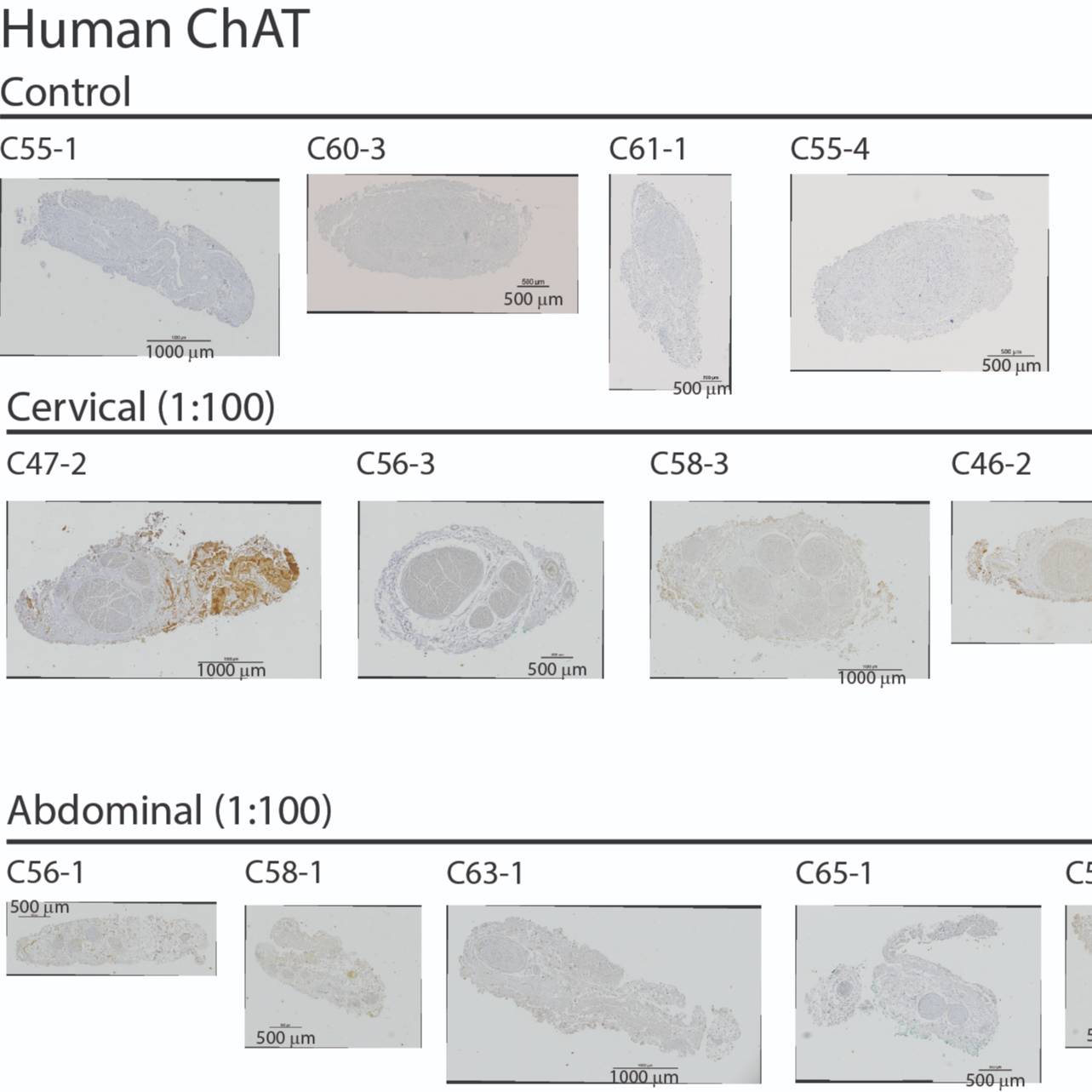
This dataset provides immunohistological images of cross sections of human vagus nerves, identifying ChAT+ and TH+ fibers.
Dataset Overview
Study Purpose: This dataset provides immunohistological images of cross sections of human vagus nerves, identifying choline acetyl transferase positive, ChAT+, and tyrosine hydroxylase positive, TH+, fibers, thereby providing information on the quantity and localization of parasympathetic and sympathetic efferent fibers within the vagus nerve.
**Data Collection:**The dataset contains image files (TIF format) for 9 left cervical and 9 anterior subdiaphragmatic human vagus nerve samples, each labeled with one of two antibodies: anti-ChAT or anti-TH, totaling ~7 GB. Section stains were interleaved, with anti-ChAT then anti-TH on successive sections.
Primary Conclusion: None stated.
Curator's Notes
Experimental Design: Slides were prepared from paraffin-embedded vagus nerve, stained with the appropriate antibodies/fluorophores and imaged. In this dataset, the nerves were stained to identify ChAT+ and TH+ nerve fibers.
Completeness: Complete.
Subjects & Samples: This dataset contains 23 samples of either the left cervical nerve or anterior subdiaphragmatic vagus nerve from 16 human subjects.
Primary vs derivative data: Primary data is a set of microscopy images in TIF format. Derivative data is formatted as JP2 (also known as JPEG 2000) in addition to TIF.
Files
1 - 0 of 0 files
About this dataset
Publishing history
Cite this dataset
Tags
References
Is Supplemented by
Ashley Ezzell, J., A. Pelot, N., A. Clissold, K., & M. Grill, W. (2019). SPARC_Duke_Grill_OT2-OD025340_VagusNerve_IHC_TH v1. https://doi.org/10.17504/protocols.io.6hehb3e
Ashley Ezzell, J., A. Pelot, N., A. Clissold, K., & M. Grill, W. (2019). SPARC_Duke_Grill_OT2-OD025340_VagusNerve_IHC_ChAT v1. https://doi.org/10.17504/protocols.io.6hfhb3n

Copyright © 2026 University of Pennsylvania. All rights reserved.