Vagus nerve stimulation mapping in swine
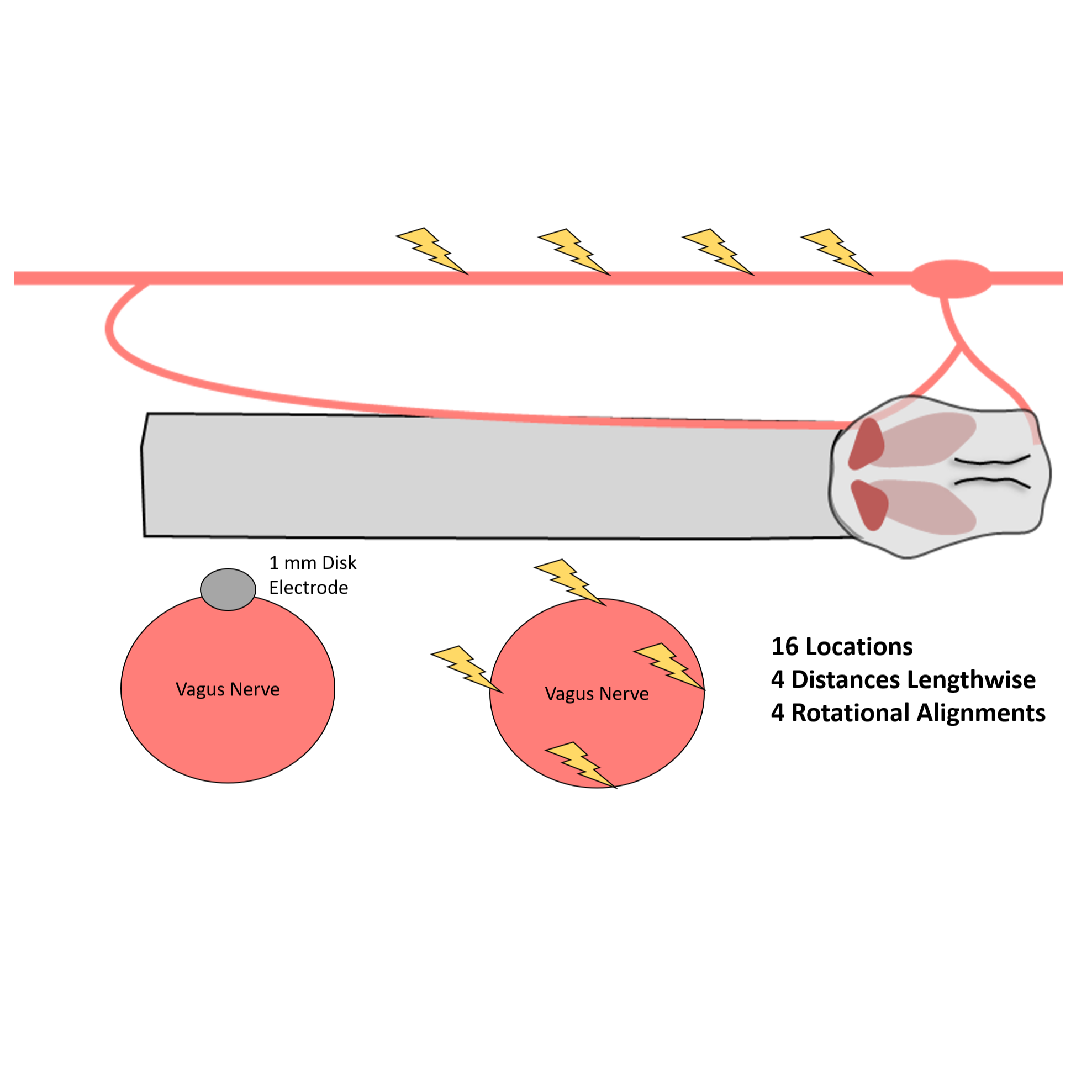
VNS evoked vagus nerve compound action potential and neck muscle electromyography data via stimulation at a 1 mm disk electrode at many locations on vagus nerve
Dataset Overview
Study Purpose: To investigate neck muscle responses to vagus nerve stimulation applied at different locations on the vagus nerve.
Data Collection: Electrophysiology and electromyography was collected using a Tucker Davis Technology system in swine in response to vagus nerve stimulation.
Primary Conclusion: The superior laryngeal branch is activated via current leakage from the stimulation electrode cuff insulation while stimulating caudal to the branching point, and superior laryngeal activation can be avoided by moving the stimulation electrode caudally away from the branch. Activation of the recurrent laryngeal branch fibers, which run under the electrode cuff at all locations in the cervical vagus, cannot be avoided via moving the stimulation electrode. The threshold for activation of the recurrent laryngeal branch fibers could be modulated by hundreds of microamperes, but EMG responses associated with recurrent laryngeal branch fiber activation were always observed before any other response (heart rate or breathing changes) associated with other fiber activation.
Experimental Design: Intrafascicular electrodes were placed within the vagus nerve to record electroneurographic (ENG) responses, and needle electrodes were placed in the vagal-innervated neck muscles to record electromyographic (EMG) responses. Bipolar stimulation was delivered at 30 Hz using symmetric biphasic pulses with 200 μs per phase and amplitudes from 50 to 3000 μA evaluated in random order. Stimulation was typically delivered for 30 s at each amplitude with at least 1 min rest between trials to allow cardiac responses to return to baseline; in some cases, stimulation was delivered for 3 s at each amplitude with at least 10 s rest due to time constraints.
Completeness: Dataset is complete.
Subjects & Samples: Male (n=3) and female (n=4) domestic pigs between 3-4 months of age were used in this study.
Primary vs derivative data: Each specific nested data folder nested under primary data (sub-) includes raw electrophysiology recordings organized by individual session subfolders. Parameters of the individual sessions can be found in the performances.xlsx file. There is no derivative data folder.
Code Availability: Source Matlab code used to analyze all the electrophysiology and electromyography data are provided in the code folder. Use the notes files in the "docs" folder to understand entries into the code. There are multiple tabs (bottom right in excel) for each notes file.
Files
1 - 0 of 0 files
About this dataset
Publishing history
Cite this dataset
Tags
References
Is Supplemented by
N Nicolai, E., & Ludwig, K. (2020). Vagus Nerve Stimulation Evoked Electroneurography and Electromyography Recordings in Swine v1. https://doi.org/10.17504/protocols.io.bkeyktfw

Copyright © 2026 University of Pennsylvania. All rights reserved.