High-throughput segmentation of rat unmyelinated axons by deep learning
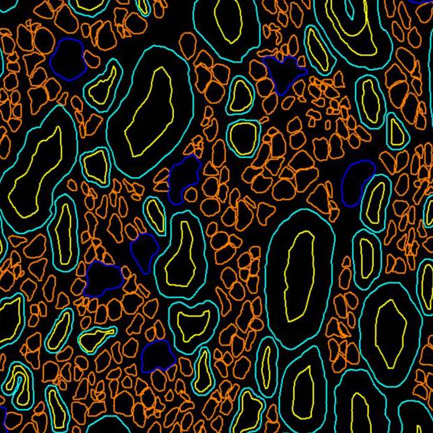
Transmission electron microscopy (TEM) images and segmentation of nerve fibers
Dataset Overview
Study Purpose: To automatically identify unmyelinated axons in nerve tissues. The manual segmentation of unmyelinated fibers, which are the majority of fibers in the nervous system, takes too much time. The correct identification of fibers is very important for the development of neuromodulation strategies.
Data Collection: Rat vagus and pelvic nerves transmission electron microscopy (TEM) images were used to create a new prototype of a high-throughput processing pipeline for automated segmentation of unmyelinated fibers.
Primary Conclusion: The new prototype saved about 80% of effort when compared with manual segmentation. This new tool will enable fast and accurate characterization of unmyelinated fibers and help to better understand the nervous system for improvement of neuromodulation strategies.
Curator's Notes
Experimental Design: Cervical, anterior, and posterior abdominal vagus nerve biopsies were obtained in both male and female rats. Immediate immersion fixation of the samples in a mixed aldehyde solution was followed by tissue processing and embedding in a plastic resin. Ultrathin sections at 70–90 nm in the transverse plane were obtained from Epon-embedded vagus nerve tissue blocks and analyzed in a transmission electron microscope (TEM) operating at 80 kV (Tecnai G2 Spirit Twin, FEI®, ThermoFisher Scientific®). Electron micrographs of regions of interest (ROI) were collected at 6500–42,000 × magnification using a Gatan Orius SC 1000B digital camera (Gatan®, Inc.), and tiling of composite EM images into montages was performed using Adobe Photoshop® (version: 21.1.3 20200508) or Image Composite Editor (ICE®, Microsoft). Data segmentation of individual myelinated and unmyelinated axons was performed manually or semi-automatically using Neurolucida® 360 (MBF Bioscience).
Completeness: This dataset is complete.
Subjects & Samples: Male (n=4) and female (n=2) Sprague-Dawley rats (RRID:RGD_737903) 49 to 68 days old were used in this study.
Primary vs derivative data: Primary data is organized by the subject ID into the sample subfolders. Each subfolder contains electron micrographs in .tif format that were obtained from representative cervical and abdominal portions of the vagus nerve and tiled. Computer-supported manual segmentation of myelinated and unmyelinated fibers in .XML format is provided for each sample. The derivative folder contains image data (JPEG2000 and OME-TIFF) that was derived from primary images (.tif). The .tif images were converted with 20:1 compression to JPEG2000 (.jpx) by MBF Bioscience for web streaming and visualization on the SPARC Data Portal. .TIF images were also converted with lossless compression to OME-TIFF (.tif) by MBF Bioscience.
Code Availability: All training, testing, and evaluation scripts are hosted on a GitHub repository at this address https://github.com/Banus/umf_unet.
Files
1 - 0 of 0 files
About this dataset
Publishing history
Cite this dataset
Tags
References
References
Havton, L. A., Biscola, N. P., Plebani, E., Rajwa, B., Shemonti, A., Jaffey, D., Powley, T. L., Keast, J. R., Lu, K.-H., & Dundar, M. (2022). High-throughput segmentation of rat unmyelinated axons by deep learning (Version 1) [Dataset]. SPARC Portal. https://doi.org/10.26275/K0MX-JCTH
Is Supplemented by
Biscola, N., & Havton, L. (2019). Nerve tissue processing for transmission electron microscopy (TEM) v1. https://doi.org/10.17504/protocols.io.xpxfmpn
Powley, T., & Jaffey, D. (2021). Collection of rat vagal tissue samples for TEM imaging v1. https://doi.org/10.17504/protocols.io.bzwcp7aw

Copyright © 2026 University of Pennsylvania. All rights reserved.