Simulations of pelvic and vagus neural interface anatomy-dependent stimulus and recording properties
This is a dataset of simulations of planar neural interface stimulation and recording properties computed for single axons and neural ensembles using ViNERS, looking at the influence of fascicle size and perineurum thickness.
Dataset Overview
Study Purpose: This hybrid computational and neuroanatomical study was undertaken to determine the influence of nerve anatomy (fascicle diameter and perineurium thickness) on the stimulation and recoding performance of planar electrode arrays using ViNERS.
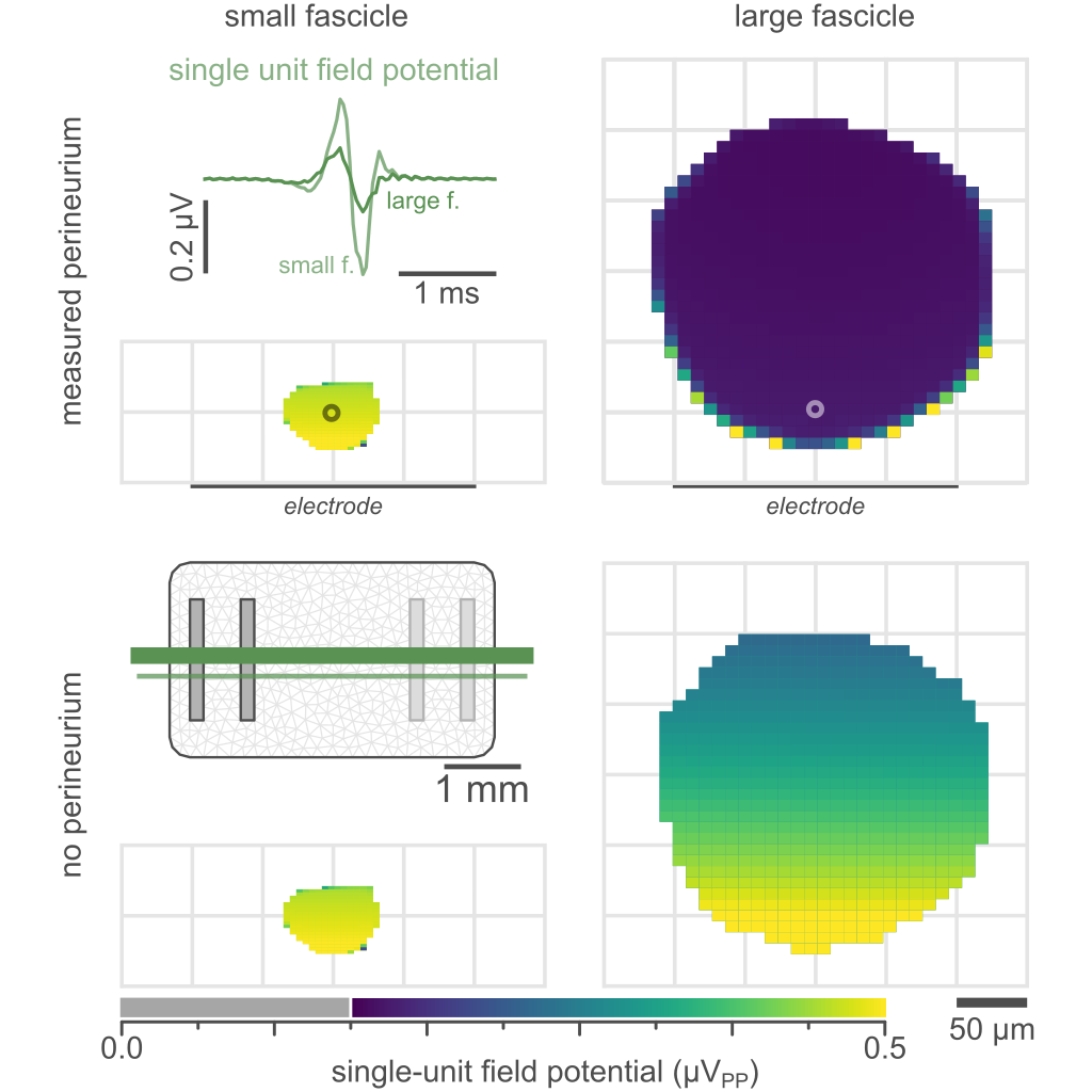
Data Collection: Single-unit field potentials (SUFPs) and single-axon extracellular stimulation thresholds were computed for myelinated and unmyelinated axons in nerve fascicles ranging from 27 to 302 µm in diameter, sampled from rat pelvic and vagus nerves. Pelvic nerve fascicles were extracted from Havton (2022) and Vagus nerve fascicles were extracted from Pelot (2020).
Electrically-evoked compound action potentials (ECAPs) and whole-nerve recordings were simulated to compare to previously recorded pelvic nerve ECAPs
Primary Conclusions: Extracellular stimulation thresholds increased and SUFP magnitudes decreased with fascicle size. Simulations of recordings from multifasciculated nerves were greater in amplitude than from single-fascicle nerves of equal area with equivalent axon-electrode separation. To generate realistic ECAPs, the fascicle geometry and the morphological diversity of axons within the nerve must be considered.
Curator's Notes
Experimental Design: A computation model.
Completeness: This dataset is a part of a larger study: ViNERS (Visceral Nerve Ensemble Recording & Stimulation) peripheral neural interface modeling environment.
Subjects & Samples: No live animals were used in this study. Values were either computed or extracted from other datasets.
Primary vs derivative data: Primary data is organized by subject ID and contains simulated values of planar neural interface stimulation and recording properties computed for single axons and neural ensembles using ViNERS.
Code Availability: Snapshot of the ViNERS code is provided in the code folder. Versioned code available at https://gitlab.unimelb.edu.au/lab-keast-osborne-release/ViNERS
Files
1 - 0 of 0 files
About this dataset
Publishing history
Cite this dataset
Tags
References
Described by
Eiber, C. D., Payne, S. C., Biscola, N. P., Havton, L. A., Keast, J. R., Osborne, P. B., & Fallon, J. B. (2021). Computational modelling of nerve stimulation and recording with peripheral visceral neural interfaces. Journal of Neural Engineering, 18(6), 066020. https://doi.org/10.1088/1741-2552/ac36e2

Copyright © 2026 University of Pennsylvania. All rights reserved.