Acute effects of efferent and afferent vagus nerve stimulation (VNS) on neural activity accessed with functional Magnetic Resonance Imaging (fMRI) in rats (Part 2)
This study aims to evaluate the effects of efferent/afferent VNS settings on neural activity in rats. Functional Magnetic Resonance Imaging (fMRI) was performed to access the whole brain neural activity.
Dataset Overview
Study Purpose: This study aims to evaluate the effects of efferent/afferent vagus nerve stimulation (VNS) on neural activity in rats.
Data Collection: Briefly, functional Magnetic Resonance Imaging (fMRI) was performed to access the whole brain neural activity. The stimulation was delivered with afferent/efferent vagotomy. The imaging session was repeated multiple times for each animal to achieve better results. The same trial was repeated six times for each animal and generate six NIfTI files for fMRI data and six matlab files for stimulation information.
Primary Conclusion: none stated
Curator's Notes
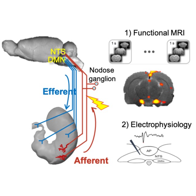
Experimental Design: The 10 rats underwent acute surgery for implantation of a bipolar cuff electrode on the left cervical vagus nerve. Experimental animals were allocated into 2 groups as follows: (i) a group of rats (N=5) received afferent VNS; (ii) the other group of rats (N=5) received efferent VNS. For the group of animals receiving afferent VNS, vagotomy was performed on the efferent side of the cuff electrode.Therefore, the stimulation only affects efferent vagus nerve. Similarly, for the group of animals receiving efferent VNS, vagotomy was performed on the afferent side of the cuff electrode, such that the stimulation only affects the afferent vagus nerve. For both groups, brain fMRI was acquired after the animal preparation. VNS was delivered during the imaging acquisition. The stimulation is the same for both group (current=0.25mA, pulse width=0.5ms, frequency=5Hz). The stimulation followed the pattern 20s-ON-40s-OFF.
Completeness: This is a part of a larger dataset.
Subjects & Samples: This study used adult Sprague-Dawley male rats (n=10) between 6-11 weeks old.
Primary vs derivative data: Primary data contains data recording of functional Magnetic Resonance Imaging (fMRI) for each animal, the fMRI data was stored in NIfTI files, and the stimulation information was stored in mat files. The size of the primary data is approximately 11 GB. Derivative data consisits of preprocessed fMRI data.
Important Notes: Every animal may experience different stimulation parameters. The information of the selected stimulation parameter can be found in the spreadsheet in each subject folder.
Files
1 - 0 of 0 files
About this dataset
Publishing history
Cite this dataset
Tags
References
Is Supplemented by
Cao, J., Lu, K.-H., Wang, X., & Liu, Z. (2020). Effects of Vagus Nerve Stimulation/Gastric Electrical Stimulation on Brain Neural Activity Assessed with Magnetic Resonance Imaging and electrophysiology v1. https://doi.org/10.17504/protocols.io.bciwiufe

Copyright © 2026 University of Pennsylvania. All rights reserved.