Effects of subcutaneous nerve stimulation on nerve sprouting in ambulatory dogs
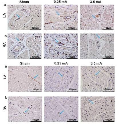
Growth-Associated Protein 43 (GAP43), tyrosine hydroxylase (TH) staining for LA, RA, LV, RV in dogs after 2 months of subcutaneous nerve stimulation (0, 0.25, 1.5, 2.5, and 3.5 mA)
Dataset Overview
Study Purpose: The rationale of the study was to test the hypothesis that subcutaneous nerve stimulation (ScNS) can remodel cardiac innervation.
Data Collection: We have harvested the tissues, processed them routinely for paraffin embedding and immunohistochemical staining followed by microscope imaging. All slides were examined manually under a BX60 microscope (Olympus, Tokyo, Japan). Five representative fields at 20X objective were selected at 4 quadrants and the center portions of the slides.
Primary Conclusion: In ambulatory dogs, low output ScNS causes cardiac nerve sprouting while high output ScNS is antiarrhythmic. Published https://doi.org/10.1016/j.hrthm.2019.02.027
Curator's Notes
Experimental Design: 22 dogs were divided into 5 different output groups for 2 months and subjected to simulation of ScNS: 0 mA (sham) (N=6), 0.25 mA (N=4), 1.5 mA (N=4), 2.5 mA (N=4) and 3.5 mA (N=4). The dogs then underwent non-survival surgery under general anesthesia. The tissues were harvested for immunostaining and terminal deoxynucleotidyl transferase dUTP nick end labeling (TUNEL) assay.
Completeness: This dataset is a part of a larger study : "Antiarrhythmic and proarrhythmic effects of subcutaneous nerve stimulation in ambulatory dogs", dataset: N:dataset:f4c832f3-3b35-4a2d-963e-6924af16cdf8
Subjects & Samples: Adult male (n=11) and female (n=11) adult dogs were used in this study.
Primary vs derivative data: Data in the primary folder is divided by the subject identification. Each subject folder contains a jpg file with microscopic images of growth-associated protein 43 (GAP43), tyrosine hydroxylase (TH) staining in the LA (left atrium), RA (right atrium), LV (left ventricle), RV (right ventricle). In total, 763 JPG images, 3.5 GB. Image data in derivative folder (JPEG2000 and OME-TIFF) was derived from primary images (.JPEG). .JPEG images were converted with lossless compression to JPEG2000 (.jp2) by MBF Bioscience for web streaming and visualization on the SPARC Data Portal. .JPEG images were also converted with lossless compression to OME-TIFF (.tif) by MBF Bioscience.
Important Notes: This dataset is currently undergoing image, segmentation, and/or scaffold curation and will be updated once this is complete.
Files
1 - 0 of 0 files
About this dataset
Publishing history
Cite this dataset
Tags
References
Described by
Wan, J., Chen, M., Yuan, Y., Wang, Z., Shen, C., Fishbein, M. C., Chen, Z., Wong, J., Grant, M. B., Everett, T. H., & Chen, P.-S. (2019). Antiarrhythmic and proarrhythmic effects of subcutaneous nerve stimulation in ambulatory dogs. Heart Rhythm, 16(8), 1251–1260. https://doi.org/10.1016/j.hrthm.2019.02.027
Is Supplemented by
not provided, P.-Sheng. C., Kusayama, T., Wan, J., Yuan, Y., Liu, X., Li, X., Shen, C., C Fishbein, M., H Everett, T., & Peng-Sheng Chen, not provided. (2021). Trichrome Staining Protocol in studies ofEffects of subcutaneous nerve stimulation on nerve sprouting in ambulatory dogs v1. https://doi.org/10.17504/protocols.io.bv94n98w

Copyright © 2026 University of Pennsylvania. All rights reserved.