3D imaging of enteric neurons in a male mouse
Confocal image projection of enteric ganglia in a male mouse
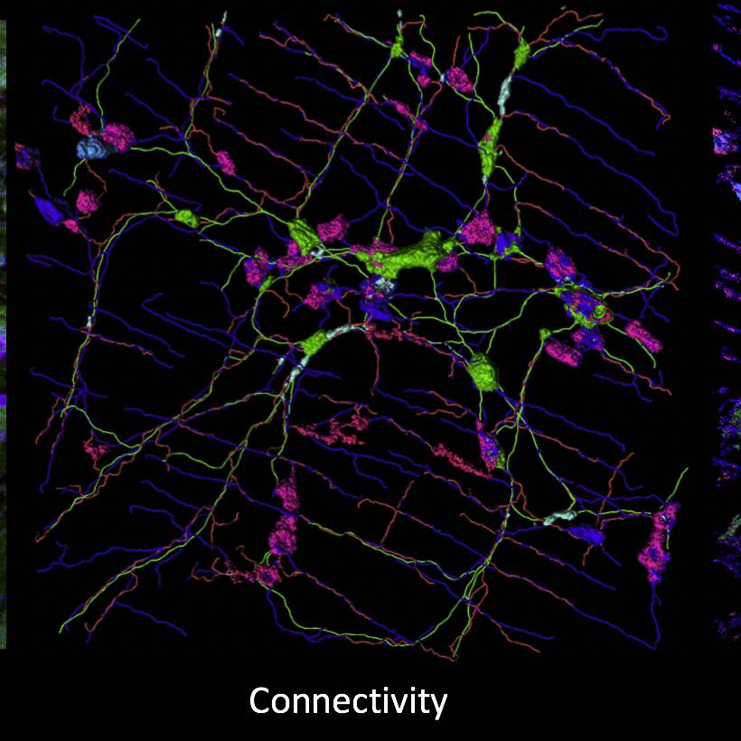
Dataset Overview
Study Purpose: To examine intra-interganglionic connectivity, neurochemical code, and morphology.
Data Collection: This dataset contains confocal projections of colonic myenteric ganglia from VIP reporter mouse.
Primary Conclusion: None stated
Curator's Notes
Experimental Design: A single VIP reporter male mouse was anesthetized, surgery performed, the distal colon dissected and fixed in PFA for 24 hours. Whole-mount stained preparations were imaged via confocal microscopy. Individual Z plane images by the channel labeling VIP (ch00 green), ChAT (ch01 red), and NOS (ch02 blue) were collected.
Completeness: This dataset is part of a larger study: "Quantitative Morphology, Transcriptomics, and Function of Enteric Neurons Expressing VIP"
Subjects & Samples: This study used one, 45 day old, male VIP reporter mouse (RRID:IMSR_JAX:031628).
Primary vs derivative data: The primary folder contains raw confocal images (.lif) collected from one sample. Individual Z plane images by the channel are also available in primary data. The derivative folder contains movies through the stack and a converted source microscopy image.
Important Notes: Primary data is stored in the proprietary Leica format. The user should open .lif – Leica Interface File first with all the needed information pertaining to the imaging session. Tif images can be opened in image J.
Files
1 - 0 of 0 files
About this dataset
Publishing history
Cite this dataset
Tags
References
Is Supplemented by
Howard, M. (2019). Wholemount Immunolabeling for GUT Samples v1. https://doi.org/10.17504/protocols.io.wr6fd9e
Described by
Nestor-Kalinoski, A., Smith-Edwards, K. M., Meerschaert, K., Margiotta, J. F., Rajwa, B., Davis, B. M., & Howard, M. J. (2022). Unique Neural Circuit Connectivity of Mouse Proximal, Middle, and Distal Colon Defines Regional Colonic Motor Patterns. Cellular and Molecular Gastroenterology and Hepatology, 13(1), 309-337.e3. https://doi.org/10.1016/j.jcmgh.2021.08.016

Copyright © 2026 University of Pennsylvania. All rights reserved.