Antibodies tested in the colon – Mouse
List of antibodies that were tested in the mouse colon. Information on source, species, dilution, and whether the test was performed on whole colonic wall with CLARITY, or whole mount preparations of submucusal and myenteric plexus.
Dataset Overview
Study Purpose: To establish a table listing antibodies that we tested and characterized to label enteric neurons and/or nerve fibers in the mouse colon. The table will serve as a reference to assess antibodies' suitability to be used in immunostaining of mouse colon enteric nervous system.
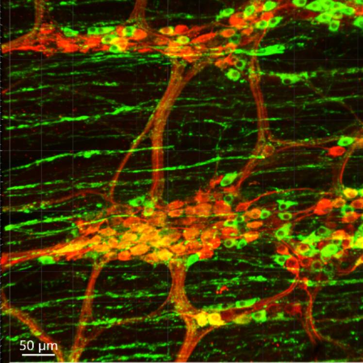
Data Collection: the murine colon samples were prepared as (1) whole mounts of the submucosal layer and myenteric and longitudinal muscle layers and (2) whole colonic wall cleared by passive CLARITY technique (PACT). Immunofluorescent methods used antibodies to label the neurons with pan-neuronal markers, neuro peptides, and classical neurotransmitter-synthesizing enzymes. A few antibodies for non-neuronal markers that play an important role in motility and inflammatory response were also tested, including the macrophage markers, glial cells, and interstitial cells of Cajal (ICC) markers. Photomicrographs were acquired in confocal microscopes. Remarks and notes about the antibodies are briefly described, and some representative figures are assembled in a file for a preview of immunofluorescent staining.
Primary Conclusion: The dataset provides information on primary antibodies tested in the mouse colon, which has been less studied than the small intestine of guinea pigs and rats. In addition, the labeling of colonic submucosal layers or whole wall processed with CLARITY was rarely reported before. The data collected herein allowed the creation of a comprehensive list of antibody labeling of colonic enteric neurons and/or nerve fibers, enteric glial cells, and interstitial cells of Cajal (ICC) in the mouse, pig, and human: SPARC Antibody Labeling Database: Colonic enteric nervous system
Curator's Notes
Experimental Design: Immunofluorescent methods were used to test the antibodies listed in the mouse colon tissues prepared as whole mounts of the submucosal and myenteric plexuses of mouse proximal and distal colon; frozen sections of the colon; or the colon with whole thickness processed by passive CLARITY technique. Photomicrographs were acquired by confocal microscopy.
Completeness: This is one of a dataset series of "Antibodies tested in the colon," which consists of three datasets: 1. human, 2. pig, and 3. mouse. Sets of "human" and "pig" are under construction.
Subjects & Samples: Adult C57BL/6J, female (n=4), and male (n=8) mice 7-12 weeks old were used in this study.
Primary vs derivative data: Primary data folder contains original confocal images of immunofluorescence of tested antibodies in the mouse colon. The images are organized in folders by subject and sample ID names, respectively. The antibodies' full names can be found in the ["abbreviations"] file in docs folder.
Important Notes: Some [special features and technical notes] of immunolabeling are remarked in an Excel file and demonstrated in a [ppt presentation] file in the "docs" folder. The antibodies and images are placed in alphabetic order. Searching the RRID online, you will find more information and references about the antibodies. The references for the non-commercial antibodies that do not have RRID are listed at the bottom of the table.
Files
0 - 0 of 0 files
About this dataset
Publishing history
Cite this dataset
Tags
References
Is Supplemented by
Wang, L., Challis, C., Liang, H., Li, S., Fowlkes, C., Sullivan, A., SR, K., & Taché, Y. (2020). Multicolor adeno-associate virus labeling and 3D digital tracing of enteric plexus in mouse proximal colon v1. https://doi.org/10.17504/protocols.io.bqavmse6

Copyright © 2025 University of Pennsylvania. All rights reserved.