Intracranial EEG Epilepsy - Study 3
Data for a patient with epilepsy obtained for clinical treatment and collected for research, consisting of a large intracranial EEG dataset (grid, strip, and depth electrodes) with multiple seizures.
Dataset Overview
The patient is a right-handed, 21-year old male who was admitted to the epilepsy monitoring unit for intracranial monitoring. The age at onset was 13 years old.
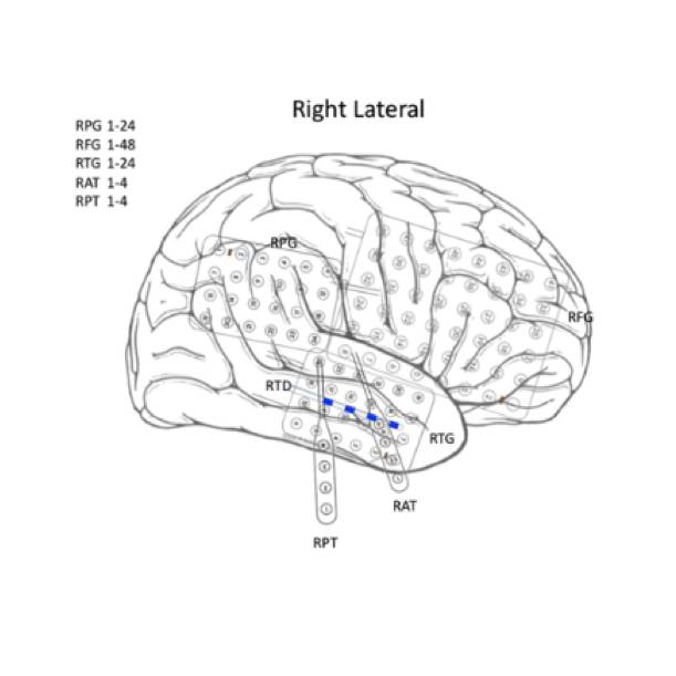
These data are from a 6 x 8 grid that was placed over the right frontal region, two 4 x 6 grids placed over the right temporal lobe and right parietal region, respectively, a 4-contact strip wrapped beneath the right anterior temporal lobe, a 4-contact strip wrapped beneath the right posterior temporal lobe, and a 4-contact depth electrode inserted in the anterior temporal region.
This is an abnormal computer-assisted prolonged intracranial EEG monitoring session due to the presence of frequent independent right frontal, right parietal and right temporal epileptiform discharges. During the monitoring session, the patient had a total of eleven seizures, including nine clinical seizures and two seizures which were subclinical. The first six seizures showed a diffuse ictal onset over the right frontal and right temporal grids. The last five seizures showed either a focal ictal onset simultaneously from the right anterior temporal strip and the posterior superior region of the right frontal grid, or focal onset from these two foci independently. These findings could be consistent with a localization-related epilepsy with seizure onset in the right frontotemporal neocortex.
The patient underwent surgery of reopening the right forntotemporoparietal craniotomy and removing the subdural grid electrodes, then a right anterior-superior frontocortical resection, a right temporal lobectomy, and a right amygdalohippocampectomy. Pathology samples of leptomeningeal and focal parenchymal lymphohistiocytic infiltrate consistent with grid/electrode placement. Mild subpial and subcortical gliosis. The patient also underwent surgery for a right mesial temporal lobe resection. Pathology samples of mesial temporal structures with focal parenchymal macrophage aggregate consistent with electrode placement. Mild subpial and subcortical gliosis.
Files
1 - 0 of 0 files
About this dataset
Publishing history
Cite this dataset
Tags

Copyright © 2026 University of Pennsylvania. All rights reserved.