Retrograde tracing of interscapular brown adipose tissue (iBAT) specific sympathetic neurons in mice - virus and reporter testing
Document and analyze the efficiency of retrograde viral transport amongst different viral constructs and mouse models
Dataset Overview
Study Purpose: The purpose of its study is to document and analyze the efficiency of retrograde viral transport amongst different viral constructs and mouse models.
Data Collection: We utilize pseudorabies virus (PRV) retrograde tracing in combination with reporter mice, iDISCO, & confocal/light-sheet microscopy to identify the location of pre- & post-ganglionic neurons as well as nerves that selectively innervate the iBAT in the mouse. Here we present tabularized cell counts of virally-labeled sympathetic chain ganglia.
Primary Conclusion: Transgenic TH-cre mice (RRID:IMSR_JAX:008601; Jackson Laboratories) crossed with reporter mice showed insufficient labeling sympathetic neurons (1-2 neurons per ganglion). In contrast, knock-in TH-IRES-cre mice (RRID:IMSR_EM:00254; B6.129X1-Thtm1(Cre)Te/Kieg; European Mouse Mutant Archive) achieved strong and consistent labeling of sympathetic neurons and nerve endings. However, during development, TH is also transiently expressed in parasympathetic precursor cells (reviewed by Howard MJ 2005, Developmental Biology, 277:271-286), so that germline transmission needs to be considered to prevent unexpected experimental outcomes. We have noted this issue in our most recent publication (Huesing C et al. 2020).
The efficiency of retrograde viral transport: a) AAV6 was most efficient for retrograde labeling of postganglionic cell bodies in sympathetic chain ganglia. AdV-cre and CAV2-cre showed substantially less labeling and were not further tested. b) Reporter expression from all viral constructs showed better visibility of RFP compared to EGFP. Cell counts of different virus tracing and reporter constructs available in submission 10. c) Overall viral expression of reporter genes, DREADDs, or ChR2 was unsatisfactory. However, AAV6-driven cre-expression was sufficient to generate robust expression in floxed-reporter mice (e.g., EGFP-L10 mice), so that following experiments were entirely performed with viral cre-expression and in floxed-animal models (DREADDflox; EGFP-L10flox; see Milestone 9).
Curator's Notes
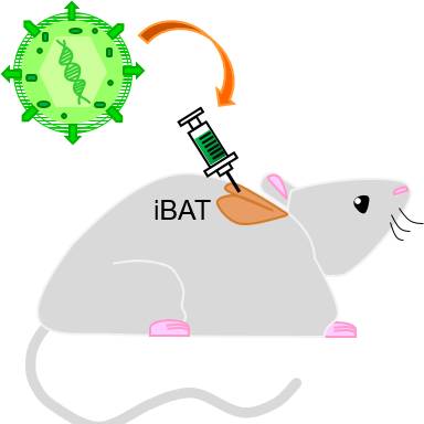
Experimental Design: The retrograde viral tracers were injected into the interscapular brown adipose tissue (iBAT) to label the axons that innervate iBAT, which have cell bodies in the sympathetic chain ganglia (SChG). After euthanasia, the peritoneal cavity is eviscerated to reveal the SChG. A stereomicroscope is used to visually confirm the extent of viral labeling within the chain ganglia. After, the samples are trimmed to only include the SChG and spinal column. Then, the iDISCO technique was performed in order to capture high-resolution images of the SChG and relevant structures via light-sheet microscopy. Finally, the images generated are used to perform cell counting on individual SChG cell bodies.
Completeness: This dataset is a part of a larger study: "Genetically-based neuro-modulation of adipose tissue functions".
Subjects & Samples: Female (n=7) and male (n=8) adult mice of mixed genetic background were used in this study.
Primary vs derivative data: Derivative data is organized as a set of excel files where summative cell counts of virally-labeled sympathetic chain ganglia cells are presented for all animals used in the study. Subject names are shown internally in the table. There is no primary data.
Files
1 - 0 of 0 files
About this dataset
Publishing history
Cite this dataset
Tags
References
Is Supplemented by
Muenzberg, H. (2020). Bilateral Adeno-associated virus (AAV) injection into interscapular brown adipose tissue v1. https://doi.org/10.17504/protocols.io.bh3tj8nn

Copyright © 2026 University of Pennsylvania. All rights reserved.