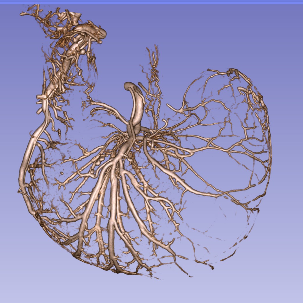
MicroCT imaging of rat stomach vasculature with Microfil MV-122
The stomach vasculature was filled at perfusion with a lead-containing silicone-based curable fluid (Microfil MV-122). Once the contrast medium was cured, the stomach was removed and fixed and subsequently imaged via microCT.
Dataset Overview
Study Purpose: As part of the stimulating peripheral activity to relieve conditions SPARC research project, we are interested in understanding how the rat stomach's dimensions vary as a function of feeding and elapsed time since feeding. Our measurements enable generation of a 4D model of the rat stomach integrating anatomical details with 2D maps of neurite distributions and other anatomical distributions. This model can then be compared with functional behavior mapping results or used to predict preferred stimulation locations for gastric electrical stimulation experiments. In particular, there is evidence that nerve fiber bundles follow the path of the vasculature; hence identifying common vasculature pathways could provide markers for electrical stimulation locations.
Data Collection: In this study, the vasculature was filled at perfusion with a lead-containing silicone-based curable fluid (Microfil MV-122). Once the contrast medium was cured, the stomach was removed, fixed and subsequently imaged via microCT. Micro-CT images were saved in DICOM format, and a subset was segmented using Vesselucida software. The data set includes 28 DICOM files collections (about 2.13 GB total when compressed), 10 XML files (about 2.8MB total), and DICOM views assembled into NIfTI-formatted files (2.74 GB).
Primary Conclusion: None stated
Curator's Notes
Experimental Design: A lead-containing silicone-based curable material was injected into the heart at perfusion and flowed through the vasculature to cure in place. Subsequent microCT imaging then provided a 3D description of the stomach vasculature.
Completeness: this dataset is complete
Subjects & Samples: Male Sprague Dawley rats (n = 28) weighing 228‐330 g were used in the study.
Primary vs derivative data: The data set includes DICOM and NRRD ("nearly raw raster data") files (in tar.bz2 directories) in bz2 compressed tape archive file for each sample organized in primary data folder by subject's name. 10 XML files (Neurolucida segmentation) and an Excel (.XLSX) spreadsheet detailing volume measurements from 3D Slicer segmentation for a subset of subjects can be found in the derivative folder.
Important Notes: This dataset is currently undergoing image registration and will be updated once this process is complete.
Files
1 - 0 of 0 files
About this dataset
Publishing history
Cite this dataset
Tags
References
Is Supplemented by
Jaffey, D., Chesney, L., & Powley, T. (2019). Micro-CT imaging of rat stomach vasculature v1. https://doi.org/10.17504/protocols.io.bafnibme

Copyright © 2026 University of Pennsylvania. All rights reserved.