Immunohistochemical classification of sensory and autonomic neurons projecting to the lower urinary tract in rats
Quantitative immunohistochemical phenotyping of peripheral sacral sensory and autonomic postganglionic neurons innervating different regions of the lower urinary tract in male and female rats.
Dataset Overview
Study Purpose: The aim of our study was to quantify functionally distinct classes of sensory and autonomic neurons that innervate the bladder body, bladder trigone, and proximal urethra, in adult male and female Sprague-Dawley rats.
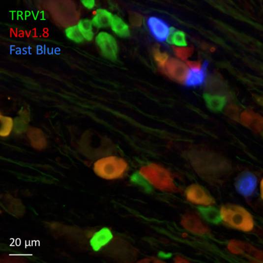
Data Collection: Microinjection of retrograde tracers (Fast Blue, FluoroGold) identified sensory and autonomic neurons projecting to each region, which were then analyzed using immunofluorescence. Lower urinary tract (LUT) dorsal root ganglion (DRG) neurons in spinal levels L6 and S1 were quantified after stratifying into nociceptive (Trpv1) and myelinated (NF200) functional classes, then further classified by their expression of peptides, sodium channels or calcium-binding proteins (n=6). Quantitation of immunohistochemically classified pelvic ganglion (PG) neurons was performed for autonomic projections to each LUT region (n=6).
Primary Conclusion: Integration of this data will enable the development of a ganglion-organ connectome for sensory and autonomic innervation of three LUT regions in male and female rats.
Curator's Notes
Experimental Design: Adult (7-9 weeks old) Sprague-Dawley rats were used for all experiments. Neural projections were identified by injecting conventional retrograde tracer dyes (fast blue, FB or Fluorogold, FG) into the target region. The site of retrograde tracer injection was: bladder (body), bladder (trigone), and urethra.
Completeness: The dataset is a part of the larger study: "Peripheral connectome of the rat lower urinary tract"
Subjects & Samples: This dataset contains immunohistochemistry data from 75 samples from 42 subjects. The samples file itself was derived from a single source file, which was a tabulated file. Please note, the structure of the source file is complex - data between and within-group factors; is nested, and has multiple counts within each sample. The researchers had to pool subsamples in some cases.
Primary vs derivative data: The primary data folder provides counts of retrograde labeled sensory neurons (tracer+) classified by immunohistochemistry. Afferent projection from sacral dorsal root ganglia [DRG (L6, S1)] to bladder (body or trigone) and urethra is presented as derived neuron counts obtained by directly counting immunostained neurons in each specimen scanned under the microscope. Individual specimen images are not part of this dataset. There is no derivative data folder. All raw counts from microscopy are tabulated and provided in the source folder.
Important Notes: The SPARC-BIDS structure required taking the master table and partitioning it into subtables, these are saved as individual files. A specimen (i.e. ganglion) was defined as a "sample", which resulted in each table having multiple rows corresponding each to a series of immunohistochemistry runs with different antibody combinations.
The documents folder contains the file "SPARC-Keast-001-Notebook", which gives a very comprehensive description of the experimental variable grouping.
Code Availability: R script to perform a connectivity analysis of neurons projecting is provided.
Files
1 - 0 of 0 files
About this dataset
Publishing history
Cite this dataset
Tags
References
Is Supplemented by
R Keast, J., & B Osborne, P. (2019). Immunohistochemical classification of sensory and autonomic neurons projecting to the lower urinary tract in rats [keast-001] v1. https://doi.org/10.17504/protocols.io.w3gfgjw

Copyright © 2026 University of Pennsylvania. All rights reserved.