Spatial distribution and morphometric characterization of vagal afferents associated with the myenteric plexus of the rat stomach
Spatial distribution and morphometric characterization of vagal afferents (specifically: intraganglionic laminar endings (IGLEs)) associated with the myenteric plexus of the rat stomach.
Dataset Overview
Study Purpose: This study investigates the correlations and causative relations between anatomical and functional characteristics of the stomach. IGLE afferents are one of the main types of vagal innervation that are associated with the stomach.
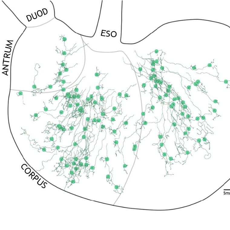
Data Collection: To characterize IGLE afferents better, rats were given injections of dextran biotin in the nodose ganglia, and, after tracer transport, stomach whole mounts were collected. Specimens were processed for avidin-biotin permanent labeling, and neurites were then digitized for morphometry and mapping using Neurolucida neural tracing software. This dataset (to date) consists of 124 XML files (~1MB to ~6 MB each) corresponding to IGLE afferents traced from Sprague-Dawley young adult male rat stomachs. Each tracing was analyzed in Neurolucida Explorer to define key neurite metrics such as area of innervation, average branch length etc. In addition, the location of the first branch point for each arbor was measured and used to construct a map of IGLE afferent locations across the stomach. In parallel activities, we are assessing the potential correlation between IGLE afferent spatial distributions and the efficacy of stimulation locations for inducing gastric motility. In addition to the digitized tracing files, the dataset also includes: (1) an Excel spreadsheet containing morphometric data, (2) a set of whole-mount photographs (jpg) used to register individual stomach contours to a standard stomach contour for generation of the 2D map; (3) a folder of 16 PNG images together with an html file that will display an interactive 2D map of neurite locations based on the associated PNG files.
Primary Conclusion: This dataset represents a detailed anatomical 2D map of the standard rat stomach as well as an interactive 2D map of neurite locations.
Curator's Notes
Experimental Design: Rats were given bilateral dextran injections into the nodose ganglia. Fourteen days after injection the rats were euthanized with either sodium pentobarbital or a mix of ketamine and xylazine. The stomach samples were removed, dissected, fixed and stained using a Vectastain Elite ABC HRP kit. A subset of the wholemount samples were counterstained with panneuronal chromogen cuprolinic blue. A third subset of the wholemount samples were counterstained against nNOS+ cells. All mounts were scanned with a Leica DMRE or a DM5500 microscope to identify afferents suitable for tracing. Neurolucida was used to trace the neurons and later perform the morphometry analyses.
Completeness: This dataset is part of a larger study with two related datasets available.
Subjects & Samples: This dataset used dorsal and ventral stomach tissue from 26 male Sprague-Dawley rats. There is a total of 34 samples.
Primary vs derivative data: The primary data consists of .xml files, while a cumulative file of the results can be found in the derivatives folder. The series of images that were used to register individual wholemount contours are contained in the source folder.
Important Notes: Most of the images that are "human" friendly, including the 2D maps are in the documents folder. The primary data folder is filled with xml flies. This dataset is has two similarly named sister datasets:, "Spatial distribution and morphometric characterization of vagal efferents associated with the myenteric plexus of the rat stomach", and “Spatial distribution and morphometric characterization of vagal afferents (intramuscular arrays (IMAs)) within the longitudinal and circular muscle layers of the rat stomach”, which use sister subjects but examine other types of vagal innervation.
Files
1 - 0 of 0 files
About this dataset
Publishing history
Cite this dataset
Tags
References
Is Supplemented by
Powley, T., Mcadams, J., & Phillips, R. (2019). High resolution labeling of vagal afferent fibers using Dextran-Biotin with counterstaining v1. https://doi.org/10.17504/protocols.io.2ipgcdn

Copyright © 2026 University of Pennsylvania. All rights reserved.